This text presents a series of excerpts from the audio piece developed by Simone C. Niquille for the Touching Thoughts project, a current cooperation between servus.at, the Department of Pathology and Molecular Pathology from the Johannes Kepler University (JKU) and the Institute of Anatomy and Cell Biology, also of the JKU.
Touching Thoughts is an art-science project that aims to explore protocols and modes of three-dimensional imaging in the field of digital pathology, and how scientific and medical knowledge and truth are generated through digital technologies.
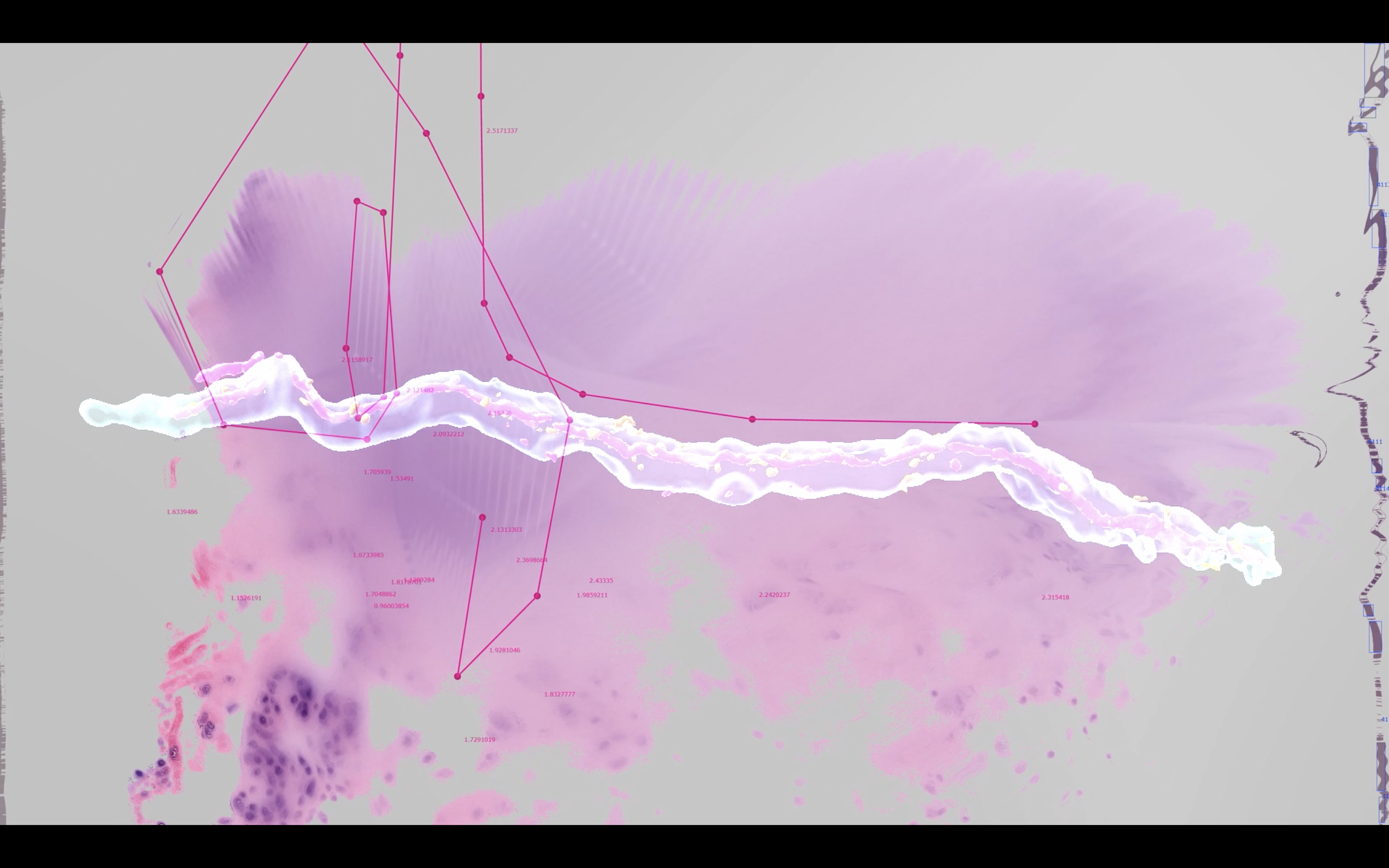
(Bild: jiawen uffline)
The collaboration involves several researchers from the Medical Faculty of JKU and invited artists Simone C. Niquille, Chaeyoung Kim & Francesco Luzzana, Bażej Kotowski, jiawen uffline and Sofia Talanti. The scientists worked with the artists to create a series of images of body tissues using confocal and light-sheet microscopy, which can produce three-dimensional microscopic and nanoscopic visualisations of brain samples and other body tissues affected by cancer or other pathologies. Such advanced imaging technologies allow researchers to reach a new order of magnitude of reality, effectively taking a medical investigation beyond the limits of physical observation. The artists involved in the project explored the images and data produced by the scientists, examining the production protocols and each focusing on selected aspects of the experiment. Chaeyoung Kim & Francesco Luzzana followed the journey of the sample and the many hands looking at it, inviting the audience to become part of this choreography of hands and gestures; jiawen uffline observes the interstices between what is the scope of observation, the cells and their components, and the constant leakage of interstitial fluids; Bażej Kotowski, inspired by the invisible rules that regulate the uncoordinated synchronicity of cellular automatisms, tested the material through transposition in the sonic realm and in volumes; Sofia Talanti delved into the representational qualities of the images, looking for the tipping points where two and three dimensions collide. Finally, Simone C. Niquille created an audio piece on the three dimensions of the human body and the technologies that try to capture its volume, interviewing scientists and documenting their goals, ideals and visions for volumetric representation.
Below is an extract from the script.
[...]
SPEAKER A
The idea for the Touching Thoughts project was to get a deeper insight into cells and tissue by capturing sample tissue in three dimensions. We want to create a novel way of looking at tissue and offer more information for analysis and diagnosis, as well as creating images that can be shown to students. We are always working with two dimensional images of three dimensional material.
INTERVIEWER
As if an architect would build a house only based on a sectional drawing, rather than the many additional dimensions a floor plan, site render and elevations offer. It would be a flat building.
SPEAKER A
Let me explain: I studied biology some years ago and I still remember the drawings of a typical cell and all its components. In a textbook you will always see a straight cut through a cell, a sectional view. It’s always visualised in a way where you have the optimal cut and you see all the specific details. But once you start to look at a real cell under the microscope, you see that reality is totally different. The image in the textbook is a very idealistic representation. Reality expands in a lot of different dimensions. Once you cut through tissue and only have one section of it to look at, you lose a lot of information. That’s why I’m especially interested in capturing a three dimensional image of a cell. I have no idea what it would look like if you could rotate a cell and move through it in three dimensions. I am curious to find out!
[...]
SPEAKER B
[...]
[Confocal microscopy] is similar to a CT scan of a human body, where you capture several images through a process that’s based on X-ray. You scan the entire human body by capturing several sections. Then you can create a 3D volume by stacking these sections in serial order to create a sort of dimensional image of the body.
INTERVIEWER
I am imagining the reverse of slicing a loaf of bread. In this case, several slices are glued back together to recreate the bread. Like watching a video in reverse.
SPEAKER B
In confocal microscopy the output is the same as a CT scan but on a microscopic level. A laser is focused on only one point, on one pixel of a cell. So you have, for example, a 0.1 millimeter thick sample tissue, which contains a lot of layers and a lot of cells. You can slice this sample tissue into several sections of 0.001 millimeter. Then you can take several scans of each layer and re-construct the tissue sample as a 3D volume. A digital re-construction of the tissue sample. By doing confocal microscopy and visualising depth, we can better look at the interaction of cells, better understand how they assemble and connect to each other and if and how they touch. Without the third dimension, it’s very hard to judge if something is touching or just really close to each other.
[...]
SPEAKER C
The slicing of the tissue samples is done by biomedical analytics assistants. They sit at a desk with a microtome, you can’t call it a machine, but it’s a structure into which you clamp the tissue sample to cut it into very very thin slices. But first the tissue sample has been soaked in formaldehyde to prevent it from decomposing and placed in paraffin, to turn it into a solid block that can be cut. Then they use the microtome, a very, very sharp knife, but it doesn’t look like a knife, more like a razor. If there’s a lot of sample tissue they can slice a few times to ultimately make a nice slice that will be mounted on the glass slide, ready to be looked at under the microscope. But if there’s just a little bit of sample tissue from a small biopsy, they have to be really very careful. It’s an art.
The biomedical analytics assistants I have spoken to, have told me that you need to feel the tissue while cutting. There is a different feel to different types of tissue even after it has been made solid with paraffin.
For example, fat tissue is squashy and you have to cut it faster than, for example, fibrotic tissue which is quite hard.
After cutting the slice it slides into a water basin. It floats on the surface because of friction. The slice is so incredibly thin. Then the glass slide is used to catch and attach the slice. It looks really, really cool.
INTERVIEWER
Beautiful, like an autumn leaf floating in a swimming pool... So, any tissue, or organ, that is removed during a surgery has to be sent to the pathology lab. Are protocols in place that guide you in creating a tissue sample? Or are you the one deciding what and where you need to cut, what tissue sample you need to create? Could you please walk us through this decision making process?
SPEAKER C
Indeed. Any part of the body that is removed, no matter how small, has to be sent to the pathology lab. It could be a small bit of skin that looks suspicious and is removed by the dermatologist or a piece of a lung that is taken out because of cancer. The body part that’s removed is placed in a container during surgery. This container is filled with a solution of formaldehyde which preserves the tissue and stops the cells from decomposing. The tissue is soaked in the solution for at least 24 hours, depending on its size.
There are cutting protocols in place for each type of organ and diagnostical question. For example, if I know that cancer could be in the tissue, there are specific ways to cut it.
We always have to think in three dimensions. Let’s say, if we take an apple and slice it in half, I get two halves with two seeds in the middle. If I were to slice it again, I see a quarter side of the apple.
Through these protocols of slicing, I can specifically cut the parts that are important for diagnostic purposes.
[...]
SPEAKER A
When we digitise the glass slides from the archive, they are always the same dimension. They are standardised at a size of around 2.5 by 7.5 centimetres. We digitize them with our scanners at 80x magnification. That produces a file of roughly 10 gigabytes. This is the largest magnification that we can get right now. We could also do smaller magnifications which produce smaller file sizes but as long as we we don’t run out of file storage space, we just use the largest magnification. The amount of slides we use for each project is always different. For projects we scan 300 slides but sometimes we scan up to 2000 slides. Scanning 2000 slides would produce somewhere around 20 terabytes of data.
Colleagues of mine developed a new cell segmentation algorithm which is pretty fast. I tested it on a couple images and it recognised roughly three million cells per slide. So if we would run this on hundreds or thousands of slides, then we would have a huge amount of individual objects that we could study, but also an enormous amount of computation and storage that is necessary. Only recently has it become possible to do this kind of work.
[...]
INTERVIEWER
When you say 3D volume, or dimensional image, I am imagining a PIXAR animated movie. But this of course is not computer generated imagery like PIXAR produces. Could you explain what three dimensional imagery means in the context of microscopical imagery?
SPEAKER B
Indeed, I often experience a kind of communication gap when I talk to people of other disciplines. To me, all images we capture for our research are three dimensional because they are depicting spatial information.
INTERVIEWER
The soviet film director Dziga Vertov said that "We cannot improve the making of our eyes, but we can endlessly perfect the camera".1 The camera, computational imaging devices and image analysis algorithms enhance our eyes and reveal patterns and information that would otherwise be invisible.
The image analysis toolset that was described earlier, which improves manual methods of tracking cancer cell's decrease and ultimately deciding on a patient's treatment, is pushing the boundaries beyond what and how the human eye can see.
Early cosmographies described the earth as a flat disk. We now know that the earth indeed is round, that cells are not flat, despite their biology textbook depiction, that the body is dimensional and that methods of visualising and capturing reality will continue to reveal, over time, new ways of seeing. While artefacts and bias are inherent, it is critical to constantly question the image and learn to read it. Over time, the various ways of seeing will stack, like slices of a bread loaf, or multiple shadows of a cylinder, towards an elaborate picture.